
IGF-1 LR3 vs IGF-1: Structural, Binding, and Half-Life Differences in Research Models
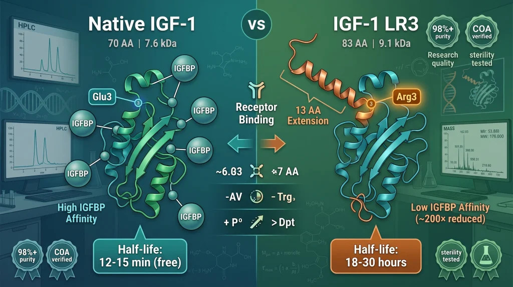
The IGF-1 LR3 vs IGF-1 comparison centers on three documented differences: an N-terminal extension, an arginine substitution at position 3, and dramatically reduced IGFBP binding affinity. These structural changes produce measurable shifts in half-life and receptor engagement across published preclinical and cell culture studies.
Understanding IGF-1 LR3 vs IGF-1: Structural Foundations
The IGF-1 LR3 vs IGF-1 comparison begins with a shared backbone: native IGF-1 (insulin-like growth factor 1) is a 70 amino acid polypeptide produced primarily in the liver in response to growth hormone signaling. Long R3 IGF-1, by contrast, is a recombinant analog engineered with two specific modifications to alter how the molecule behaves in IGF-binding-protein-rich environments such as serum and cell culture media. As described in foundational research indexed on PubMed (NCBI), these structural changes produce a variant with markedly different pharmacokinetic and binding properties from the native peptide.
Native IGF-1 circulates almost entirely bound to six IGF-binding proteins (IGFBPs), which regulate its bioavailability, transport, and tissue access. Long R3 IGF-1 was specifically engineered to evade most IGFBP binding, which is why researchers studying receptor-mediated effects independent of binding-protein interference often select the analog over the native peptide. Understanding the structural origin of this difference is the foundation of the IGF-1 LR3 vs IGF-1 decision in research design.
The Long R3 Modification: N-Terminal Extension and Arginine Substitution
Long R3 IGF-1 differs from native IGF-1 by two engineered changes that, together, define the analog’s behavior in receptor and binding-protein assays. Both modifications were introduced to address specific limitations researchers encountered when using recombinant human insulin-like growth factor in IGFBP-rich systems.
The 13 Amino Acid N-Terminal Extension
The “L” in LR3 refers to a 13 amino acid extension added to the N-terminus of the IGF-1 sequence. This extension is derived from a methionyl porcine growth hormone fragment and is fused to the natural IGF-1 sequence during recombinant expression. The added residues do not appear to participate directly in IGF-1 receptor binding, but they contribute to the analog’s increased stability and altered binding profile, as described in protein engineering literature indexed by the U.S. National Library of Medicine.
The Arg3 Substitution
The “R3” portion of the name refers to the substitution of glutamic acid at position 3 of the native IGF-1 sequence with arginine. This single-residue change has an outsized effect on the analog’s behavior: it dramatically reduces affinity for all six IGFBPs while leaving binding to the IGF-1 receptor largely intact. The arginine substitution is therefore the principal driver of the IGFBP-evasion property that distinguishes the IGF-1 LR3 vs IGF-1 profile.
Research Insight: Why Both Modifications Matter
The N-terminal extension and the Arg3 substitution act through different mechanisms. The extension contributes to structural stability and resistance to proteolysis, while the Arg3 substitution specifically disrupts IGFBP interaction. Researchers who require a variant with reduced binding-protein interference and improved stability in long-duration cell culture experiments often turn to the LR3 form. View research-grade IGF-1 LR3 specifications →
Sequence Snapshot
The combined effect of these two changes is a peptide of approximately 83 amino acids (70 native + 13 N-terminal extension) with a single residue substitution at the third position of the native portion. While the molecular weight increases from roughly 7.6 kDa for native IGF-1 to approximately 9.1 kDa for Long R3 IGF-1, the IGF-1 receptor recognition surface remains essentially intact.
IGFBP Binding Affinity: The Defining Difference in IGF-1 LR3 vs IGF-1
In the IGF-1 LR3 vs IGF-1 comparison, no parameter shows a larger documented gap than IGFBP affinity. Native IGF-1 binds the six IGFBPs with high affinity, and in serum more than 95% of IGF-1 circulates in ternary complex with IGFBP-3 and the acid-labile subunit. Long R3 IGF-1, by design, binds IGFBPs roughly two to three orders of magnitude less avidly, freeing the analog to interact with the IGF-1 receptor without competing for binding-protein partitioning.
How IGFBPs Modulate Native IGF-1
The IGFBP system serves as a reservoir, transporter, and regulator of IGF-1 in vivo. Binding proteins extend native IGF-1’s half-life from minutes (free peptide) to hours (complexed form) and gate its tissue access. In cell culture media containing serum, secreted IGFBPs can sequester added recombinant human insulin-like growth factor, reducing the effective concentration interacting with the IGF-1 receptor. This is a well-documented confound in growth factor research and is reviewed in literature available through the PubMed Central archive.
Why LR3 Evades the System
The Arg3 substitution disrupts the binding interface between IGF-1 and the IGFBPs. Because Long R3 IGF-1 binds these proteins with very low affinity, far less of the added analog is sequestered. In practical cell culture terms, a lower dose of Long R3 IGF-1 can produce a receptor-engagement signal equivalent to a much higher dose of native IGF-1. The IGF-1 LR3 vs IGF-1 affinity gap is the single most cited reason researchers select the analog for IGFBP-rich experimental systems.
Cell Culture Implications
For researchers using serum-supplemented media, the IGFBP-evasion property of LR3 translates into more reproducible dose-response curves and reduced lot-to-lot variability from serum-derived binding proteins. This is one reason recombinant Long R3 IGF-1 is widely listed in cell culture media formulations for studies on satellite cell activation, skeletal muscle hypertrophy models, and protein synthesis assays. The functional consequence is that researchers can interrogate IGF-1 receptor signaling pathways with less confounding from upstream binding-protein dynamics.
Half-Life and Receptor Engagement: Comparative Research Data
Differences in IGFBP binding directly produce differences in measured half-life. The IGF-1 LR3 vs IGF-1 half-life comparison varies depending on whether researchers measure free peptide, complexed peptide, or total immunoreactive species, but the qualitative pattern is consistent across studies.
Native IGF-1 Pharmacokinetics
Free native IGF-1 has a very short circulating half-life, typically reported in the range of 10 to 15 minutes in unmodified plasma. When complexed with IGFBP-3 and the acid-labile subunit, the apparent half-life extends to roughly 12 to 15 hours in rodent and primate studies, reflecting the protective role of the binding-protein system. This dynamic is foundational to understanding how the recombinant human insulin-like growth factor behaves in vivo versus in vitro.
Long R3 IGF-1 Pharmacokinetics
Because Long R3 IGF-1 binds IGFBPs only weakly, it does not enter the long-lived ternary complex. Yet, its measured half-life remains longer than that of free native IGF-1, generally reported between 18 and 30 hours in rodent models. This counterintuitive result is attributed to the stabilizing effect of the N-terminal extension and resistance to certain proteolytic pathways, rather than to binding-protein protection. A useful summary of comparative pharmacokinetic data is available through NIH-indexed research repositories.
Receptor Affinity Considerations
Long R3 IGF-1 retains nearly full affinity for the IGF-1 receptor, although reported values vary slightly across studies. Its affinity for the insulin receptor remains low, similar to that of native IGF-1, which is relevant to researchers concerned about cross-talk between insulin-like growth factor 1 signaling and insulin pathway activity. Reported cross-reactivity at the insulin receptor for both molecules is roughly 1% to 2% of insulin’s own affinity.
⚠️ 5 Important Research Considerations for IGF-1 LR3 vs IGF-1
These considerations matter for designing valid experiments and interpreting findings across published growth factor research.
- Variant selection drives data interpretation: Findings from Long R3 IGF-1 experiments cannot be directly extrapolated to native IGF-1 outcomes due to the IGFBP-evasion difference.
- Media composition changes the comparison: In serum-free media with no IGFBP background, the IGF-1 LR3 vs IGF-1 dose response converges more closely than in serum-supplemented systems.
- Receptor specificity should be confirmed: Both variants engage the IGF-1 receptor but show negligible insulin-receptor activation; assay design should confirm receptor identity in mixed-receptor models.
- No human therapeutic equivalence: Neither IGF-1 LR3 nor native IGF-1 is FDA-approved for human therapeutic use in the way the LR3 analog is sometimes informally discussed; LR3 in particular is research-grade only.
- Lot-to-lot purity matters: Reported potency differences across studies often reflect purity variance, aggregation state, or endotoxin contamination rather than intrinsic mechanism differences.
Consequence Examples
Issue: Using native IGF-1 in serum-rich media at low dose → Research Impact: Most peptide sequestered by IGFBPs, blunting observed receptor activation.
Issue: Cross-comparing LR3 and native potency without controlling for IGFBP content → Research Impact: Apparent potency difference reflects binding-protein dynamics, not intrinsic receptor affinity.
Issue: Sourcing peptide without a current Certificate of Analysis → Research Impact: Reproducibility issues attributed to biology may actually trace to purity or aggregation.
Published reviews consistently note that variant-selection rationale must be reported alongside dose, vehicle, and media composition for findings to be reproducible across laboratories.
Sourcing research-grade peptides with verified purity documentation reduces these variables significantly. View Phoenix Peptide Labs quality standards →
✅ What the Research Shows About IGF-1 LR3 vs IGF-1
These principles are validated across more than a decade of peer-reviewed growth factor research and are referenced in literature indexed on PubMed and PubMed Central.
Research backing for these principles spans growth factor biochemistry, recombinant protein engineering, and cell culture methodology, with key sources reviewed in NCBI archives.
Researchers who apply these principles consistently report more reproducible dose-response curves, lower batch-to-batch variability, and greater confidence interpreting IGF-1 receptor signaling pathways.
How to Source High-Quality IGF-1 LR3 for Your Research
Quick Answer
Sourcing research-grade Long R3 IGF-1 requires verifying purity by HPLC (typically ≥98%), confirming sequence identity by mass spectrometry, reviewing a current Certificate of Analysis, and confirming endotoxin and sterility testing. US-manufactured material with batch-level documentation supports the most reproducible cell culture and preclinical outcomes in IGF-1 LR3 vs IGF-1 comparison studies.
The quality of the source peptide is often the single largest controllable variable in growth factor research. The factors below outline what to verify before any IGF-1 LR3 vs IGF-1 experiment.
1. HPLC Purity Verification
High-performance liquid chromatography (HPLC) is the standard analytical method for confirming the purity of recombinant Long R3 IGF-1. Research-grade material typically reports ≥98% purity by HPLC, with the chromatogram included in the Certificate of Analysis.
Lower-purity material can carry truncated species, oxidation products, or aggregation byproducts that alter dose-response in unpredictable ways. Researchers should request the HPLC trace alongside the percent-purity number, since the trace itself reveals impurity profiles a single value cannot.
2. Mass Spectrometry Identity Confirmation
Mass spectrometry confirms that the molecular weight of the material matches the expected ~9.1 kDa for Long R3 IGF-1. Identity confirmation distinguishes correctly expressed LR3 from native IGF-1 or mis-spliced variants, which is critical for any IGF-1 LR3 vs IGF-1 comparison study.
Look for documentation indicating both intact-mass analysis and, where available, peptide-mapping confirmation of the Arg3 substitution and the N-terminal extension.
3. Endotoxin and Sterility Documentation
For cell culture work, endotoxin contamination can confound results by triggering inflammatory signaling pathways unrelated to growth factor mechanism. A research-grade Certificate of Analysis should report endotoxin levels (typically by LAL assay) and a sterility statement for the lyophilized product.
The FDA’s guidance on bacterial endotoxin testing describes the analytical standards underlying these measurements, even though research-grade material is not regulated for human use.
Ready to source research-grade IGF-1 LR3 with full documentation? Shop Phoenix Peptide Labs’ verified peptide catalog →
4. US Manufacturing and Chain of Custody
US-manufactured peptides offer the strongest documentation chain, including raw material sourcing, lot traceability, and adherence to consistent quality protocols. For research that may be published, peer-reviewed, or replicated across laboratories, traceability of the source material is part of the methodological transparency reviewers increasingly expect.
Independent third-party COA verification adds a further layer of confidence, particularly for studies where the IGF-1 LR3 vs IGF-1 comparison itself is a published outcome.
5. Storage, Handling, and Reconstitution Specifications
Long R3 IGF-1 is supplied lyophilized and is stable at -20°C or colder for extended periods. After reconstitution in bacteriostatic water or an appropriate research vehicle, working solutions are typically stable for shorter windows under refrigeration, with aliquoting recommended to minimize freeze-thaw cycles.
A reliable supplier will provide handling and reconstitution guidance specific to their product form, since impurity profiles and excipient choices can affect post-reconstitution stability.
6. Lot-Level Bioactivity Reporting
Some suppliers report bioactivity per lot via standardized cell-based assays. This is not universal in the research peptide market but adds confidence for laboratories running standardized dose-response curves across multiple shipments.
When bioactivity is not reported per lot, researchers can establish an internal calibration curve using a known-good reference lot for ongoing IGF-1 LR3 vs IGF-1 comparison work.
7. Documentation and Compliance Records
Beyond the COA, qualified researchers should expect access to material safety data sheets, lot records, and a clear statement that the material is intended for research use only and is not approved for human or veterinary therapeutic use.
Suppliers that restrict sales to qualified researchers and laboratories operate within the appropriate regulatory framework for research chemicals, as outlined in general guidance from the National Institutes of Health on research material standards.
IGF-1 LR3 vs IGF-1 Specification Comparison Table
The table below summarizes the parameters most commonly cited in published comparisons. Values reflect ranges reported across multiple peer-reviewed studies and are not exhaustive.
| Parameter | Native IGF-1 | Long R3 IGF-1 (LR3) | Research Significance |
|---|---|---|---|
| Amino acid length | 70 | 83 (70 + 13 N-terminal) | Structural footprint |
| Molecular weight | ~7.6 kDa | ~9.1 kDa | Identity confirmation by MS |
| Position 3 residue | Glutamic acid (Glu) | Arginine (Arg) | Drives IGFBP evasion |
| IGFBP-3 affinity | High (Kd in pM range) | ~200× reduced | Key cell culture variable |
| IGF-1 receptor affinity | Reference (~1.0) | ~0.7–1.0 of native | Functional preservation |
| Insulin receptor affinity | ~1–2% of insulin | ~1–2% of insulin | Pathway specificity |
| Reported half-life (rodent models) | ~12–15 min free / ~12–15 hr complexed | ~18–30 hr | Bioactivity duration |
| Regulatory status | Research / approved analogs exist | Research use only, not FDA-approved | Compliance framing |
Values compiled from peer-reviewed literature indexed on PubMed and PubMed Central, 2017–2024. Ranges reflect inter-study variation.
Shop Research-Grade IGF-1 LR3 →Phoenix Peptide Labs Quality Standards
Every batch of research-grade IGF-1 LR3 sold through Phoenix Peptide Labs is supported by:
- HPLC purity verification (≥98% target specification)
- Mass spectrometry identity confirmation
- Third-party Certificate of Analysis per lot
- Endotoxin and sterility testing documentation
- US-manufactured with lot-level traceability
Review documentation and order with Certificate of Analysis →
Who Uses IGF-1 LR3 vs IGF-1 in Research?
The IGF-1 LR3 vs IGF-1 comparison appears across cell biology, growth factor signaling, and recombinant protein research. Below are representative scenarios.
The Cell Culture Researcher
Working with skeletal muscle cell lines or satellite cell models in serum-containing media. Wants to interrogate IGF-1 receptor signaling without IGFBP confounding.
The Graduate Researcher Starting a Comparison Study
Designing an experiment that directly compares native IGF-1 and Long R3 variants. Needs verified purity for both molecules and reliable documentation for thesis methodology.
The Research Team Scaling an Experiment
Running a multi-cohort preclinical model where peptide consistency across shipments is essential. Lot-level documentation and reproducibility are non-negotiable.
Have questions about your research needs? Contact our team →
Frequently Asked Questions: IGF-1 LR3 vs IGF-1
Long R3 IGF-1 differs from native IGF-1 by a 13 amino acid N-terminal extension and an arginine substitution at position 3, which together reduce IGFBP binding affinity by roughly 200-fold while preserving IGF-1 receptor engagement.
The structural changes produce a longer reported half-life in preclinical models and reduced sensitivity to binding-protein sequestration in cell culture systems. As reviewed in literature on PubMed, this is the principal mechanistic distinction reported across IGF-1 LR3 vs IGF-1 studies.
For sourcing questions or documentation requests, contact our team.
Long R3 IGF-1’s extended half-life reflects the combined effect of the N-terminal extension’s structural stability and reduced clearance pathways available in the absence of strong IGFBP binding.
While native IGF-1 in complex with IGFBP-3 can also show extended half-life, free LR3 maintains bioactivity longer than free native IGF-1 because the extension confers intrinsic proteolytic resistance. Reported rodent-model half-lives for LR3 typically fall in the 18 to 30 hour range, summarized across studies indexed by NCBI.
Replacing glutamic acid at position 3 with arginine introduces a positive charge at a residue that normally contributes to the IGFBP binding interface, disrupting the contact and reducing binding affinity across all six IGFBPs.
This single-residue change explains most of the IGFBP-evasion property of Long R3 IGF-1. Researchers studying recombinant human insulin-like growth factor variants frequently cite the Arg3 substitution as the engineered feature responsible for the analog’s distinct cell culture behavior.
Research-grade material is typically specified at ≥98% purity by HPLC, with mass spectrometry identity confirmation and lot-level documentation.
Lower purity introduces truncated species, oxidation byproducts, or aggregation that can alter dose-response and confound IGF-1 LR3 vs IGF-1 comparison data. Endotoxin and sterility documentation are additionally important for cell culture applications, as outlined in general guidance from the FDA bacterial endotoxin guidance referenced for research material standards.
Need technical specifications or purity documentation? Contact Phoenix Peptide Labs directly →
Lyophilized Long R3 IGF-1 is typically stored at -20°C or colder, with reconstituted working stocks aliquoted to minimize freeze-thaw cycles.
Reconstitution typically uses bacteriostatic water or a research-appropriate vehicle, with post-reconstitution stability windows specified by the supplier. Maintaining cold chain during shipping and storage is essential to preserving peptide integrity for IGF-1 LR3 vs IGF-1 experiments.
Research-grade material carries documented HPLC purity, mass spectrometry identity confirmation, endotoxin and sterility testing, and a current Certificate of Analysis per lot.
Lower-grade material may lack one or more of these signals, which introduces variability that can mask or mimic biological effects. For studies depending on the IGF-1 LR3 vs IGF-1 comparison itself, documentation depth is a methodological requirement.
Most published Long R3 IGF-1 data come from in vitro cell culture and rodent preclinical models, with limited direct comparison to native IGF-1 in identical experimental conditions.
Heterogeneity in media composition, vehicle, and dose ranges across studies makes meta-analysis challenging, and the IGFBP-evasion property means LR3 findings cannot be straightforwardly extrapolated to systems where native IGF-1 dynamics dominate.
No. IGF-1 LR3 is not FDA-approved for human or veterinary therapeutic use. It is sold strictly for laboratory and research applications and is restricted to qualified researchers and laboratories.
Some native IGF-1 analogs have specific approved indications in narrow clinical settings, but Long R3 IGF-1 is not among them and is consistently classified as research use only across regulatory references including the FDA.